23+ Color Ultrasound Images
Ultrasound images are displayed in either 2D 3D or 4D which is 3D in motion. These ultrasound images of a young asymptomatic adult female patient reveal multiple calcific plaques of the common carotid arteries of both sides.

Ultrasound And Color Doppler Pulse Clinic
Ultrasound images of flow whether color flow or spectral Doppler are essentially obtained from measurements of movement.
Color ultrasound images. Colour Doppler displays the direction and flows velocities of the blood superimposed on the cross-sectional image. If youre 12 weeks along in the pregnancy you may be able to make out your babys head and if youre 20 weeks along you may even see the spine heart feet and eyes. Reflected ultrasound waves have altered properties eg altered amplitude.
This image from the lab of Professor Kevin Parker shows the color imaging possible with his H-scan format compared to traditional ultrasound imaging around it showing only shades of gray. Blue means the blood is streaming away from the transducer and red means the blood is moving towards the transducer Note. Ultrasonography pregnant ultrasound ultrasonography woman thyroid pregnancy male ultrasound pregnant woman having ultrasound ultrasound image thyroid and pregnancy ultrasound table ultrasound breast.
Mary McMahon In order to perform a color Doppler ultrasound the ultrasound machine must be capable of producing color Doppler imaging. Introduction This paper was based on the idea that the human visual system is more responsive to color than binary or monochrome images. Rated 500 out of 5 based on 1 customer rating.
Color Doppler ultrasound image shows a grade 3 varicocele of the left side. Structures are enhanced. The ultrasound probe transducer is placed over the carotid artery top.
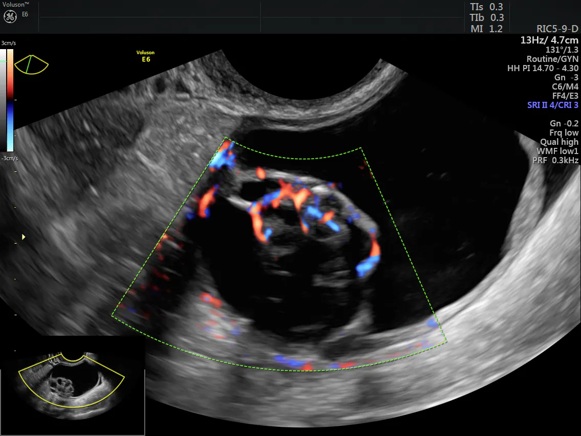
In children before 5 years of age ovaries have a volume of less than 1 cm. Advancements in ultrasound technology include three-dimensional 3-D ultrasound that formats the sound wave data into 3-D images. Ultrasound and color Doppler images of Dystrophic calcification of carotid arteries.
The pampiniform veins of the left side measure almost 44 mm on valsalva maneuver. 1 customer review 1900000 1749000. The next two images correspond to a grayscale obstetric ultrasound left and its matching color post-processed image right.
The right scrotum is also affected and show a grade 3 varicocele affecting a. Color Atlas of Ultrasound Anatomy Second Edition presents a systematic step-by-step introduction to normal sectional anatomy of the abdominal and pelvic organs and thyroid gland essential for recognizing the anatomic landmarks and variations seen on ultrasound. Ultrasound images of flow whether color flow or spectral Doppler are essentially obtained from measurements of movement.
62939 ultrasound stock photos vectors and illustrations are available royalty-free. Ultrasound Color Processing is the result of programmed algorithms applied to ultrasound pictures and for different purposes. See ultrasound stock video clips.
ISonic High Quality Images Color Ultrasound Scanner. In ultrasound scanners a series of pulses is transmitted to detect movement of blood. Other links to color or gray scale images will be included here later.
Color in diagnostic ultrasound may indicate different things. With this technique it became possible to directly observe blood flow within the heart. Breakthrough adds new color to ultrasound.
The use of color flow Doppler CFD or color Doppler imaging CDI or simply color Doppler sonography allows the visualization of flow direction and velocity within a user defined areaA region of interest is defined by the sonographer and the Doppler shifts of returning ultrasound waves within are color-coded based on average velocity and direction. A smaller color box gives a higher frame rate and better image. ISonic Wide band multi-frequency Imaging Processing Imaging optimization and Compound enhance technology touch pad and keyboard high quality images and.
Blue and red does not necessarily mean low-oxygen and high-oxygen blood. A similar process would be used for Ultrasound Color Pre-processing. The advent of color Doppler was a breakthrough in medical ultrasound.
Echoes from stationary tissue are the same from pulse to pulse. Its convenient double-page format with more than 250 image quartets. Calcific lesions of carotid arteries.
Color Doppler ultrasound is a special ultrasound technique that evaluates blood flow through a blood vessel including the bodys major arteries and veins in the abdomen arms legs and neck. Affects the quality of the image. We wanted to experiment with the importance of pseudo-color.
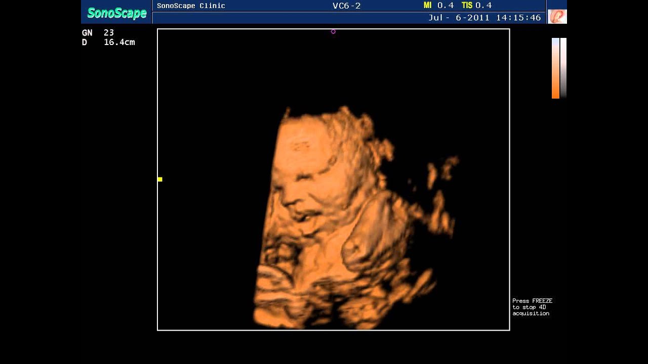
Four-dimensional 4-D ultrasound is 3-D ultrasound in motion. Seroma hematoma abscess tumors hernias foreign bodies like bullets glass. To read an ultrasound picture look for white spots on the image to see solid tissues like bones and dark spots on the image to see fluid-filled tissues like the amniotic fluid in the uterus.
Answer 1 of 8. The images obtained transrectally are quite similar to those obtained transvaginally. A PRF of 45 to 60 cmsec is optimal for the great arteries and a range of 10 to 20 cmsec is optimal for venous structures.
Such visualization is achieved by color-encoding Doppler information and displaying the colors as an overlay on the 2D image of the heart. A color ultrasound image bottom left shows blood flow the red color in the image in the carotid artery. This is exploited to give the reflected sound waves based on their amplitude different nuances on the ultrasound image.
The tissues in the ultrasound image are drawn with varying shades of one color usually gray. Delivery in 3 Days. These images are examples of pathology I detect with sonograms.
This device images frequency changes in the colors blue and red. In ultrasound scanners a series of pulses is transmitted to detect movement of blood. Color Doppler ultrasound is a medical imaging technique which is used to provide visualization of the bloodflow using color processing to add color to the image so that a doctor or care provider can clearly see what is happening inside the body.
Echoes from stationary tissue are the same from pulse to pulse. Image enhancement Ultrasound images Pseudo-Color Transforms Filters 1. This case of recurrence of varicocele of the left scrotum.
One of the applications of Doppler ultrasound is color Doppler fig. 6971 likes 34 talking about this. A gallery of ultrasound images including B mode 3D 4D ultrasound and color Doppler sonography.
Color Doppler ultrasound video clips of varicocele. First of all and most often this is the so-called color flow mode for blood flow visualization which is usually represented by gradations of two colors most often gradations of blue and red but there may be other c. Gold orange and yellow.
The first links in each row here correspond to ultrasound color post-processed images. Power Doppler and Colour Doppler Ultrasound Power Doppler is a form that is more sensitive to slow flow but is described as one color eg. Best of all there is a short description.
Before sexarche ovaries can be visualized by transabdominal ultrasound using the full bladder technique or by transrectal approach using the transvaginal probe. Velocity scale or PRF pulse repetition frequency.

4d Sonoscape S8 Color Doppler Ultrasound Real Baby Youtube
Using Color Doppler To Classify Ovarian Tumors Empowered Women S Health
China Obstetric Color Doppler Sonography Manufacturers And Suppliers Factory Pricelist Sunbright

China Classical Portable Color Ultrasound Scanner Photos Pictures Made In China Com

One Way To Use Your Colour Doppler When Performing A Canine Pregnancy Scan Animal Ultrasound Association
Color Doppler Ultrasound Images Shows Blood Flow Signals In The Wall Download Scientific Diagram

Breakthrough Adds New Color To Ultrasound Newscenter
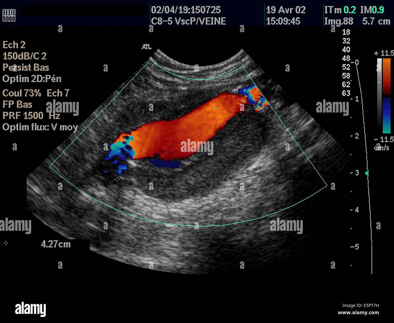
Color Doppler Ultrasound Scan Of An Aneurysm Of The Abdominal Aorta Showing Circulating Lumen Of The Artery Orange Stock Photo Alamy
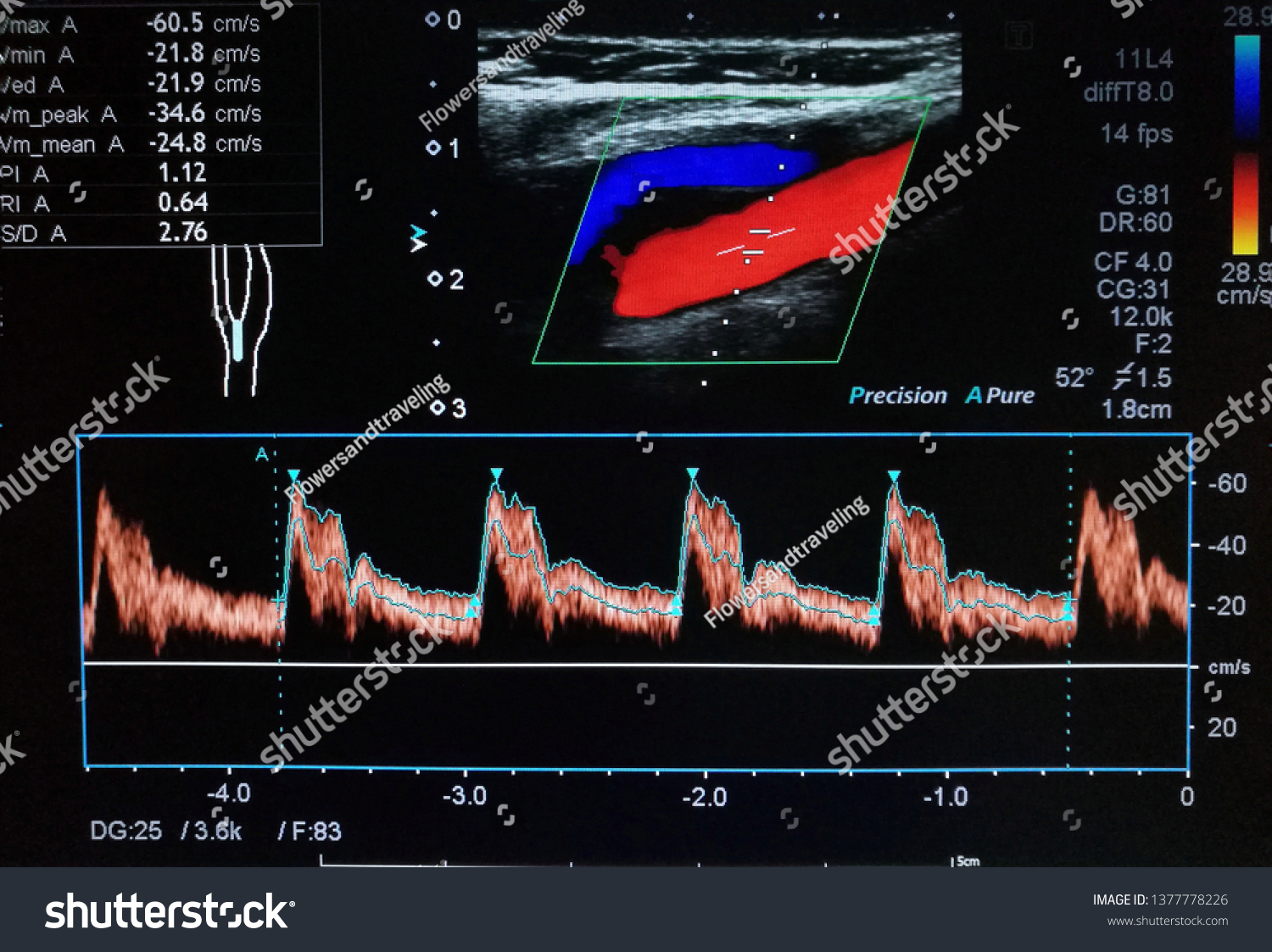
Color Doppler Ultrasound Imaging Normal Common Stock Photo Edit Now 1377778226


Comments
Post a Comment